The MULE Team has worked in several areas of research over the years. This page highlights some of the major accomplishments and successes the MULE Team has achieved.
EXAMPLES OF MAJOR ACCOMPLISHMENTS:
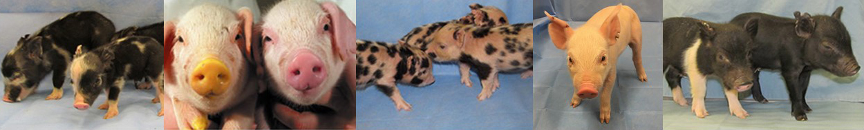
1. Porcine Reproductive and Respiratory Syndrome Virus (PRRSV) devastates pig populations worldwide. The economic costs are $6 Million dollars per day in North America and Europe with even larger losses reported in Asia. PRRSV vaccines are not very effective. The literature suggested two proteins were independently responsible for viral infection. Knockout of the first candidate gene (SIGLEC1) was started. Due to the dearth of funding, it took 10 years to create animals to challenge. Unfortunately, those pigs were not resistant to the virus. As the project to test the second gene (CD163) began, we adapted CRISPR/Cas9 technology for use in pigs and in less than 6 months, edited founder animals were produced. These animals are healthy, reproduce, grow well, and are resistant to challenge by North American and European strains of the virus. A patent has been issued to the University of Missouri and the technology is licensed to Genus plc. Genus is currently seeking FDA approval for the PRRSV resistant pigs. This particular gene edit addresses several issues:
A) Animal welfare as animals suffer when sick,
B) Sustainability by increasing production efficiency,
C) Food security as infection results in unpredictable decreases in the supply of pork, and
D) The psychological, emotional, and financial costs to family farms upon loss of their pigs.
2. Gene Knockouts for major diseases include:
ANTXR1 to make the pigs resistant to Senecavirus A,
ANPEP to make pigs resistant to Transmissible Gastroenteritis,
CD1D to study the importance of NK T-Cells, and
TMPRSS2 for potential resistance to a variety of viruses.
3. The tools for genetic engineering are having a major impact on the way researchers are now able to answer fundamental questions. For example, Rod Geisert has an interest in maternal recognition of pregnancy. Proteins that are candidates for this process have been studied for the past 30 years. Now, the tools are available to begin to answer unequivocally some of these questions. Recently, a proof of principle was published (Whyte et al ’18) confirming that IL1B2 is responsible for a major morphological remodeling that occurs in the embryo. Follow-up experiments with CYP19A1 knockout are changing the dogma regarding embryo/sow communication.
EXAMPLES OF RECENT SUCCESSES:
Somatic Cell Nuclear Transfer. To improve efficiency of somatic cell nuclear transfer (SCNT), it is necessary to modify differentiated donor cells to become more amendable for reprogramming by the oocyte cytoplasm. A key feature that distinguishes somatic/differentiated cells from embryonic/undifferentiated cells is cellular metabolism, with somatic cells using oxidative phosphorylation (OXPHOS) while embryonic cells utilize glycolysis. Inducing metabolic reprogramming in donor cells could improve SCNT efficiency by priming cells to become more embryonic in nature prior to the SCNT procedure. Hypoxia inducible factor 1-α (HIF1- α), a transcription factor that allows cell survival in low oxygen, promotes a metabolic switch from OXPHOS to glycolysis. We hypothesized that chemically stabilizing HIF1- α in donor cells by the use of hypoxia mimetic cobalt chloride (CoCl2) would promote this metabolic switch in donor cells and subsequently improve SCNT efficiency. Donor cell treatment with 100 µM CoCl2 for 24 hours prior to SCNT up-regulated the mRNA abundance of glycolytic enzymes, improved SCNT blastocyst development rate and quality, and also affected gene expression in the blastocysts. After transfer of blastocysts created from CoCl2 treated donor cells to surrogates, healthy cloned piglets were produced. Therefore, stabilization of HIF1- α in donor cells by CoCl2 treatment is a simple, economical way to improve SCNT efficiency.
A simple and fast PCR-based protocol for sex determination in pigs. Advances in genome editing tools have reduced barriers to the creation of animal models. Due to their anatomical and physiological similarities to humans, there has been a growing need for pig models to study human diseases, for xenotransplantation and translational research. The ability to determine the sex of genetically modified embryos, cells or fetuses is beneficial for every project involving the production of transgenic animals. This strategy can improve the time-efficiency and lower the production costs. Thus, we developed a simple and fast PCR-based protocol for sex determination in pigs by using a unique primer set to amplify either the DDX3X or DDX3Y gene. The sex was 100% correctly assigned when tail genomic DNA, Day-35 fetus and hair samples from pigs were used. For both blastocysts and oocytes (88% and 96% of efficacy, respectively) the unidentified samples were potentially due to human error. Our assay worked for domestic sheep (Ovis aries), American bison (Bison bison), and European cattle (Bos taurus) samples and by in silico analysis, we confirmed X-Y amplicon length polymorphisms for the DDX3 gene in 12 other mammalian species. This PCR protocol for determining sex in pig tissues and cells showed to be simple, specific, highly reproducible and less time consuming as well as an important tool for other livestock species and wildlife studies.